Difference between revisions of "Lobule VIIIA"
Jump to navigation
Jump to search
| (9 intermediate revisions by 3 users not shown) | |||
| Line 1: | Line 1: | ||
| − | <meta name="title" content="Lobule VIIIA"/> | + | <!-- <meta name="title" content="Lobule VIIIA"/> --> |
| − | {{cer nav}} | + | {{cer nav|lobule=true}} |
| + | __NOTOC__ | ||
{{h2|Lobule VIIIA}} | {{h2|Lobule VIIIA}} | ||
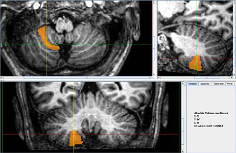
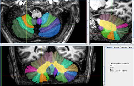
| + | *Location: Lower regions of the cerebellum, extends from midline laterally | ||
| + | *Description: Best to identify and label the boundary for VIIIB before VIIIA as the lower boundary can be difficult to identify otherwise | ||
| + | **The fissure between this lobule and Lobule VIIA Crus I is easily identified in the midsagittal slice. | ||
| + | *This lobule usually consists of one or two white matter branches and its second boundary is characterized by the exclusion of VIIIB which has a distinct curve. | ||
| + | <gallery widths="500" heights="300"> | ||
| + | image:lobuleVIIIA.jpg | ||
| + | image:lobuleVIIIA_all.jpg | ||
| + | </gallery> | ||
| − | + | <gallery widths="300" heights="200"> | |
| − | + | image:AnonKwijibo45_Lobe8A_certain.jpg|''Figure 1'' : Clear and easy example of VIIIA | |
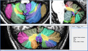
| − | + | image:AnonKwijibo46_8A.jpg|''Figure 2'' : The boundary between VIIIA and VIIB was difficult in this scan and was finally determined using the preperventure fissure in the sagittal view. | |
| − | + | image:AnonKwijibo50_8A.jpg|''Figure 3'' : Sometimes 8A has two distinct branches. | |
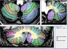
| − | + | image:AnonKwijibo56_8a_curve.jpg|''Figure 4'' : 8a wraps around 8b in an unusual way. | |
| − | + | </gallery> | |
| − | |||
| − | |||
| − | |||
| Line 20: | Line 26: | ||
{{DEFAULTSORT:{{PAGENAME}}}} | {{DEFAULTSORT:{{PAGENAME}}}} | ||
[[Category:IACL]] | [[Category:IACL]] | ||
| + | [[Category:IACL Cerebellum Pages]] | ||
Latest revision as of 00:58, 3 July 2022
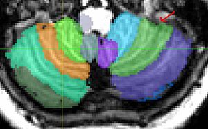
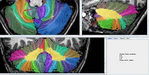
Lobule VIIIA
- Location: Lower regions of the cerebellum, extends from midline laterally
- Description: Best to identify and label the boundary for VIIIB before VIIIA as the lower boundary can be difficult to identify otherwise
- The fissure between this lobule and Lobule VIIA Crus I is easily identified in the midsagittal slice.
- This lobule usually consists of one or two white matter branches and its second boundary is characterized by the exclusion of VIIIB which has a distinct curve.
